
IMAT Expert Guide: Human Anatomy and Physiology
🌟 I. Introduction to Human Anatomy and Physiology for IMAT
A. Purpose of this Guide
This guide offers a focused and simplified overview of human anatomy and physiology, specifically tailored to the requirements of the International Medical Admissions Test (IMAT). The content emphasizes key concepts, structures, and functions that are frequently tested. Biology, which encompasses human physiology, is a critical section of the IMAT, contributing significantly to the overall score.1 A solid understanding of the topics presented here is therefore essential for success.
B. General Organization of the Human Body
The human body exhibits a remarkable hierarchical organization. The most basic structural and functional unit is the cell. Groups of similar cells performing a common function form tissues (e.g., muscle tissue, nervous tissue). Different types of tissues are organized into organs (e.g., the heart, stomach, brain), each with specific functions. Finally, organs work together in organ systems (e.g., the digestive system, circulatory system) to carry out complex physiological processes necessary for life.
It is crucial to recognize that these organ systems do not operate in isolation. They are intricately interconnected and interdependent, constantly communicating and coordinating their activities to maintain the body's overall balance, a state known as homeostasis. For instance, the digestive system breaks down food, but it relies on the circulatory system to transport absorbed nutrients to all cells, the nervous system for control of muscle contractions and secretions, and the endocrine system for hormonal regulation of digestive processes.2 Understanding these interconnections provides a more holistic view of human physiology and is vital for tackling IMAT questions that may integrate concepts across multiple systems.
😋 II. The Digestive System
A. Overview and Primary Functions
The digestive system is responsible for processing ingested food through several stages: ingestion (taking food in), digestion (breaking down large food molecules into smaller, absorbable units), absorption (passage of digested nutrients into the blood and lymph), and egestion or elimination (removal of undigested waste materials).4 Its primary importance lies in converting complex carbohydrates, proteins, and fats into simpler molecules that the body can use for energy, growth, and repair.4
B. Key Structures and Organs
The digestive system consists of the alimentary canal (a continuous tube) and accessory organs that aid in digestion.
1. Alimentary Canal
Mouth (Oral Cavity): The entry point for food.
- Mechanical digestion: Achieved by mastication (chewing), which breaks food into smaller pieces, increasing its surface area.
- Chemical digestion: Begins here with saliva, produced by salivary glands. Saliva contains the enzyme salivary amylase, which initiates the breakdown of starches (carbohydrates) into simpler sugars.4 Food is mixed with saliva to form a soft, moist ball called a bolus.
Pharynx (Throat): A common passageway for both food (to the esophagus) and air (to the larynx). The epiglottis, a flap of cartilage, covers the opening of the larynx (glottis) during swallowing, preventing food from entering the trachea (windpipe).7
Esophagus: A muscular tube that connects the pharynx to the stomach. It transports the bolus via peristalsis, which are rhythmic, wave-like muscular contractions of its walls.4
Stomach: A J-shaped, muscular organ that serves as a temporary storage site for food (can hold 2-4 liters) and continues both mechanical and chemical digestion.4
- Mechanical digestion: The stomach's muscular walls churn and mix the bolus with gastric juices, forming a semi-liquid mixture called chyme.
- Chemical digestion: Gastric glands in the stomach lining secrete gastric juice, which contains:
- Hydrochloric acid (HCl): Creates a highly acidic environment (pH 1.5-2.5) that denatures proteins (unfolding them for easier enzyme access) and kills most ingested bacteria.4
- Pepsinogen: An inactive enzyme precursor that is converted to the active enzyme pepsin by HCl. Pepsin begins the digestion of proteins into smaller polypeptides.4
- The stomach has folds called rugae that allow it to expand and increase surface area.4 It is separated from the esophagus by the cardiac sphincter (lower esophageal sphincter) and from the small intestine by the pyloric sphincter.8
📸 Image Description: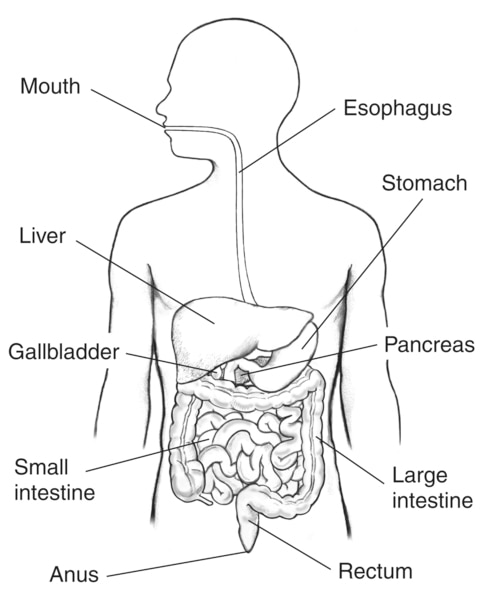
Small Intestine: The longest part of the alimentary canal and the primary site for chemical digestion and nutrient absorption. It has three sections:
- Duodenum: The first, shortest section (about 25 cm). It receives chyme from the stomach, along with bile from the liver/gallbladder and pancreatic juice from the pancreas.5
- Jejunum and Ileum: The remaining sections where most digestion is completed and the majority of nutrients (amino acids, monosaccharides, fatty acids, glycerol, vitamins, minerals) are absorbed into the bloodstream or lymphatic system. The inner surface is highly folded into circular folds, villi (finger-like projections), and microvilli (microscopic projections on epithelial cells), which vastly increase the surface area available for absorption.
Large Intestine (Colon): Shorter but wider than the small intestine.
- Its main functions are to absorb water and electrolytes (salts) from the remaining indigestible food matter and to form and store feces.5
- It houses a large population of bacteria (gut flora) that can break down some undigested material and synthesize certain vitamins, such as vitamin K and some B vitamins.5
- It consists of the cecum (with the appendix attached), colon (ascending, transverse, descending, and sigmoid portions), rectum, and anal canal.
Rectum and Anus: The rectum stores feces prior to defecation. The anus is the terminal opening through which feces are eliminated from the body.6
2. Accessory Digestive Organs
These organs are not part of the alimentary canal but produce secretions essential for digestion.
- Salivary Glands: (Parotid, submandibular, sublingual glands) Produce saliva, which moistens food, contains salivary amylase, and has some antibacterial properties.5
- Liver: The largest internal organ with many functions, including:
- Producing bile, an alkaline fluid that aids in fat digestion by emulsifying fats (breaking large fat globules into smaller droplets, increasing surface area for lipase action).5
- Detoxifying harmful substances.
- Metabolizing carbohydrates, lipids, and proteins.
- Storing glycogen, vitamins (A, D, E, K, B12), and minerals (iron).
- Gallbladder: A small organ located beneath the liver that stores and concentrates bile produced by the liver. It releases bile into the duodenum when fatty food enters.5
- Pancreas: Located behind the stomach, it has both exocrine and endocrine functions.
- Exocrine function: Produces pancreatic juice, which contains a variety of digestive enzymes (pancreatic amylase, lipases, proteases like trypsinogen and chymotrypsinogen) and bicarbonate ions (to neutralize acidic chyme from the stomach). Pancreatic juice is secreted into the duodenum.5
- Endocrine function: Produces hormones like insulin and glucagon, which regulate blood sugar levels (discussed in the Endocrine System section).2 The pancreas demonstrates how an organ can serve multiple, distinct physiological roles, linking digestion with the body's overall metabolic control.
C. Process of Digestion and Absorption
Mechanical Digestion: The physical breakdown of food into smaller pieces without changing its chemical nature. This includes chewing in the mouth, churning in the stomach, and peristalsis moving food along the tract.5
Chemical Digestion: The enzymatic breakdown of large, complex food molecules (macromolecules) into smaller, simpler molecules that can be absorbed.5
- Carbohydrates (e.g., starch, glycogen) are broken down into monosaccharides (e.g., glucose, fructose, galactose) by amylases and disaccharidases.
- Proteins are broken down into amino acids by proteases (e.g., pepsin, trypsin, chymotrypsin, peptidases).
- Fats (Lipids) are broken down into fatty acids and glycerol (or monoglycerides) by lipases, with the aid of bile for emulsification.
Absorption: The process by which the small, digested nutrient molecules pass from the lumen of the small intestine across the intestinal epithelium into the blood capillaries (for monosaccharides, amino acids, water-soluble vitamins, minerals) or lymphatic capillaries (lacteals, for fatty acids, glycerol, fat-soluble vitamins).5 Most absorption occurs in the jejunum and ileum of the small intestine.
The efficiency of absorption in the small intestine is a direct consequence of its structural adaptations. The presence of circular folds, villi, and microvilli dramatically increases the internal surface area of the small intestine by many hundreds of times. This vast surface area ensures maximum contact between the digested food particles and the absorptive cells, facilitating rapid and efficient uptake of nutrients. This principle of maximizing surface area for exchange is a common theme in biological systems.
Table 1: Key Digestive Enzymes
| Enzyme | Source (Organ/Gland) | Substrate | Product(s) |
|---|---|---|---|
| Salivary Amylase | Salivary Glands | Starch | Maltose, smaller polysaccharides |
| Pepsin | Stomach (Gastric Glands) | Proteins | Smaller polypeptides |
| Pancreatic Amylase | Pancreas | Starch | Maltose, smaller polysaccharides |
| Trypsin | Pancreas | Proteins | Smaller polypeptides, amino acids |
| Chymotrypsin | Pancreas | Proteins | Smaller polypeptides, amino acids |
| Pancreatic Lipase | Pancreas | Triglycerides (Fats) | Fatty acids, monoglycerides, glycerol |
| Intestinal Peptidases | Small Intestine (Brush border) | Polypeptides | Dipeptides, amino acids |
| Intestinal Disaccharidases (e.g., Maltase, Sucrase, Lactase) | Small Intestine (Brush border) | Disaccharides (Maltose, Sucrose, Lactose) | Monosaccharides (Glucose, Fructose, Galactose) |
🫁 III. The Respiratory System
A. Overview and Primary Functions
The primary function of the respiratory system is gas exchange: it facilitates the intake of oxygen (O₂) from the atmosphere into the body and the elimination of carbon dioxide (CO₂), a waste product of cellular metabolism, from the body.7 This process is essential for cellular respiration, the metabolic pathway that cells use to convert nutrients (like glucose) into adenosine triphosphate (ATP), the energy currency of the cell.
B. Key Structures and Organs
The respiratory system consists of a series of passages that conduct air to and from the lungs, and the lungs themselves where gas exchange occurs.
- Nose/Nasal Cavity & Mouth: These are the primary entry points for air. The nasal cavity is specialized to warm, humidify, and filter incoming air. Hairs (vibrissae) at the entrance trap larger particles, while mucus secreted by the lining traps smaller particles and pathogens. Cilia, tiny hair-like projections on the epithelial cells, beat rhythmically to move this mucus towards the pharynx to be swallowed or expelled.7
- Pharynx (Throat): A muscular funnel that serves as a common passageway for air (from the nasal cavity/mouth to the larynx) and food (from the mouth to the esophagus).7
- Larynx (Voice Box): A cartilaginous structure located between the pharynx and the trachea. It contains the vocal cords (vocal folds), which vibrate as air passes over them, producing sound. The epiglottis, a flap of cartilage at the superior end of the larynx, closes over the glottis (opening of the larynx) during swallowing to prevent food and liquids from entering the lower respiratory tract.7
- Trachea (Windpipe): A flexible tube, about 10-12 cm long, extending from the larynx into the thoracic cavity, where it divides into the two primary bronchi. Its walls are supported by C-shaped rings of hyaline cartilage, which prevent the trachea from collapsing and keep the airway open. The trachea is lined with ciliated pseudostratified columnar epithelium, which produces mucus (the "mucociliary escalator") that traps debris and pathogens and sweeps them upwards towards the pharynx.7
- Bronchi (singular: Bronchus): The trachea bifurcates into the right and left primary bronchi, each entering a lung. These bronchi further divide into smaller secondary (lobar) bronchi (three in the right lung, two in the left), then into tertiary (segmental) bronchi, and continue to branch into progressively smaller airways.7 Like the trachea, larger bronchi are supported by cartilage.
- Bronchioles: The smallest conducting airways, branching from the tertiary bronchi. They lack cartilage but have smooth muscle in their walls, allowing their diameter to be regulated (bronchodilation and bronchoconstriction). The terminal bronchioles lead to the respiratory bronchioles, which then lead to the alveoli.7
- Lungs: A pair of large, spongy, cone-shaped organs located in the thoracic cavity, one on each side of the heart. They are protected by the rib cage and surrounded by a double-layered serous membrane called the pleura (visceral pleura adheres to the lung surface, parietal pleura lines the thoracic cavity wall; the pleural cavity between them contains a thin layer of pleural fluid that reduces friction during breathing).7 The lungs contain the extensive network of bronchi, bronchioles, and alveoli.
- Alveoli (singular: Alveolus): Tiny, thin-walled air sacs, resembling clusters of grapes, found at the ends of the bronchioles. They are the functional units of the lungs where gas exchange occurs. There are hundreds of millions of alveoli in the lungs, providing an immense surface area (about 70-100 square meters, roughly the size of a tennis court) for efficient diffusion of gases.7 Each alveolus is surrounded by a dense network of pulmonary capillaries.
- Diaphragm: A large, dome-shaped sheet of skeletal muscle located at the base of the thoracic cavity, separating it from the abdominal cavity. It is the primary muscle of respiration.7
📸 Image Description:
C. Mechanism of Breathing (Ventilation)
Breathing, or pulmonary ventilation, is the mechanical process of moving air into and out of the lungs. It involves two phases: inhalation and exhalation, driven by pressure differences between the atmosphere and the lungs, created by changes in thoracic volume.
Inhalation (Inspiration): An active process that draws air into the lungs.
- The diaphragm contracts and flattens (moves downwards).
- The external intercostal muscles (located between the ribs) contract, pulling the rib cage upwards and outwards.
- These actions increase the volume of the thoracic cavity.
- As the thoracic volume increases, the lungs expand (due to pleural linkage), and the pressure inside the lungs (intrapulmonary pressure) decreases to below atmospheric pressure.
- Air flows into the lungs, moving from an area of higher pressure (atmosphere) to lower pressure (lungs).7
Exhalation (Expiration): Typically a passive process at rest, relying on the elastic recoil of the lungs and chest wall.
- The diaphragm relaxes and returns to its dome shape (moves upwards).
- The external intercostal muscles relax, and the rib cage moves downwards and inwards.
- These actions decrease the volume of the thoracic cavity.
- The elastic recoil of the lungs causes them to decrease in volume, increasing the intrapulmonary pressure to above atmospheric pressure.
- Air flows out of the lungs, moving from an area of higher pressure (lungs) to lower pressure (atmosphere).7
Forced exhalation (e.g., during exercise or coughing) is an active process involving the contraction of internal intercostal muscles and abdominal muscles, which further decreases thoracic volume and expels air more forcefully.
D. Gas Exchange
The critical function of gas exchange occurs in the alveoli by diffusion across the respiratory membrane. This membrane is extremely thin (about 0.5 micrometers) and consists of the squamous epithelial cells of the alveolar wall, the squamous endothelial cells of the capillary wall, and their fused basement membranes.
Oxygen (O₂) Exchange: The partial pressure of oxygen (PO₂) is higher in the alveolar air (inhaled air) than in the deoxygenated blood arriving in the pulmonary capillaries. Therefore, O₂ diffuses from the alveoli, across the respiratory membrane, into the blood. Here, most of it binds to hemoglobin within red blood cells to be transported to body tissues.
Carbon Dioxide (CO₂) Exchange: The partial pressure of carbon dioxide (PCO₂) is higher in the deoxygenated blood (carrying waste from tissues) than in the alveolar air. Therefore, CO₂ diffuses from the blood, across the respiratory membrane, into the alveoli to be expelled from the body during exhalation.7
The efficiency of this gas exchange is paramount and is directly related to the structural design of the lungs. The millions of thin-walled alveoli, each surrounded by a dense capillary network, provide an enormous surface area for diffusion.7 According to Fick's Law of Diffusion, the rate of gas transfer is directly proportional to the surface area available for diffusion and the concentration (or partial pressure) gradient of the gas, and inversely proportional to the thickness of the barrier it must cross. The respiratory system is exquisitely adapted to maximize these factors: a vast surface area and an extremely thin diffusion barrier, ensuring rapid and efficient uptake of oxygen and removal of carbon dioxide.
Furthermore, the respiratory tract possesses several protective mechanisms to keep the delicate alveoli free from debris and pathogens. Nasal hairs filter large particles. The mucous lining of the airways, produced by goblet cells, traps smaller particles and microorganisms. The cilia lining much of the conducting zone (trachea, bronchi) beat in a coordinated fashion, creating the "mucociliary escalator" that constantly sweeps this mucus and trapped material upwards towards the pharynx, where it can be swallowed or coughed out.7 The C-shaped cartilaginous rings in the trachea and bronchi also play a protective role by preventing these major airways from collapsing, thus ensuring a patent (open) pathway for air flow.7
❤️ IV. The Circulatory System (Cardiovascular System)
A. Overview and Primary Functions
The circulatory system, also known as the cardiovascular system, is the body's primary transport network. Its main functions include:
- Transporting oxygen from the lungs and nutrients from the digestive system to all body cells.
- Removing waste products, such as carbon dioxide (to the lungs) and urea (to the kidneys), from cells.
- Transporting hormones from endocrine glands to their target organs.
- Helping to regulate body temperature by distributing heat.
- Defending the body against disease through the actions of white blood cells and antibodies circulating in the blood.12
The system comprises three main components: the heart (a muscular pump), blood vessels (a network of tubes), and blood (the transport medium).
B. The Heart: Structure and Function
The heart is a remarkable, cone-shaped muscular organ, composed primarily of specialized cardiac muscle. It is located in the mediastinum (the central compartment of the thoracic cavity), between the lungs, and tilted slightly so that about two-thirds of its mass lies to the left of the body's midline.13 Its main function is to pump blood continuously throughout the body.
Four Chambers
The heart is a four-chambered organ, effectively acting as a double pump.
- Right Atrium: Receives deoxygenated blood returning from the systemic circulation (rest of the body) via two large veins: the superior vena cava (from upper body) and the inferior vena cava (from lower body).13
- Right Ventricle: Receives deoxygenated blood from the right atrium and pumps it to the lungs via the pulmonary artery for gas exchange.13
- Left Atrium: Receives oxygenated blood returning from the lungs via the pulmonary veins.13
- Left Ventricle: Receives oxygenated blood from the left atrium and pumps it into the aorta, the largest artery in the body, for distribution to the systemic circulation. The wall of the left ventricle is significantly thicker and more muscular than that of the right ventricle because it must generate much higher pressure to pump blood throughout the entire body, whereas the right ventricle only pumps blood to the nearby lungs.13
Valves
To ensure unidirectional blood flow and prevent backflow, the heart has four valves:
- Atrioventricular (AV) Valves: Located between the atria and ventricles.
- Tricuspid Valve: Between the right atrium and right ventricle. It has three cusps (flaps).14
- Mitral Valve (Bicuspid Valve): Between the left atrium and left ventricle. It has two cusps.14
- Semilunar Valves: Located at the exit of each ventricle, where arteries leave the heart.
- Pulmonary Valve: Between the right ventricle and the pulmonary artery.14
- Aortic Valve: Between the left ventricle and the aorta.14
Heart Wall Layers
- Epicardium: The outermost layer, also known as the visceral layer of the serous pericardium (the sac surrounding the heart).
- Myocardium: The thick, middle layer composed of cardiac muscle tissue. This is the contractile layer responsible for pumping blood.13
- Endocardium: The innermost layer, a thin sheet of endothelium that lines the heart chambers and covers the valves, providing a smooth surface to reduce friction.13
Cardiac Cycle
The sequence of mechanical and electrical events that occurs during one heartbeat. It consists of two main phases:
- Systole: The period of ventricular contraction and blood ejection.
- Diastole: The period of ventricular relaxation and blood filling.
📸 Image Description:
C. Blood Vessels
Blood vessels form a closed network of tubes that transport blood throughout the body. There are three main types:
- Arteries: Carry blood away from the heart. Most arteries carry oxygenated blood (the exception is the pulmonary artery, which carries deoxygenated blood from the right ventricle to the lungs). Arteries have thick, elastic, and muscular walls to withstand the high pressure of blood pumped from the ventricles.12 The largest artery is the aorta. Arteries branch into progressively smaller vessels called arterioles, which regulate blood flow into capillary beds.
- Veins: Carry blood towards the heart. Most veins carry deoxygenated blood (the exception is the pulmonary veins, which carry oxygenated blood from the lungs to the left atrium). Veins have thinner walls and larger lumens (internal diameters) than corresponding arteries, and they operate under lower pressure.12 Many veins, especially those in the limbs, contain valves that prevent the backflow of blood, aiding its return to the heart against gravity. Small vessels called venules collect blood from capillaries and merge to form larger veins. The largest veins are the superior and inferior vena cavae.
- Capillaries: Microscopic blood vessels that connect arterioles to venules. Their walls are extremely thin, consisting of a single layer of endothelial cells. This thinness facilitates the efficient exchange of gases (O₂, CO₂), nutrients, and waste products between the blood and the interstitial fluid surrounding body cells.12 Capillaries form extensive networks called capillary beds throughout body tissues.
D. Blood Circulation
The human circulatory system is a double circulatory system, meaning blood passes through the heart twice during one complete circuit of the body. It consists of two main circuits:
- Pulmonary Circuit: This circuit transports deoxygenated blood from the right side of the heart to the lungs, where it picks up oxygen and releases carbon dioxide, and then returns oxygenated blood to the left side of the heart.12
Path: Right Ventricle → Pulmonary Artery → Lungs (capillaries surrounding alveoli for gas exchange) → Pulmonary Veins → Left Atrium. - Systemic Circuit: This circuit transports oxygenated blood from the left side of the heart to all other tissues and organs of the body, where it delivers oxygen and nutrients and picks up carbon dioxide and waste products, and then returns deoxygenated blood to the right side of the heart.12
Path: Left Ventricle → Aorta → Systemic Arteries → Arterioles → Capillaries in body tissues (for exchange) → Venules → Systemic Veins → Superior and Inferior Vena Cavae → Right Atrium. - Coronary Circuit: A specialized part of the systemic circuit that supplies oxygenated blood to the heart muscle (myocardium) itself, via the coronary arteries, and returns deoxygenated blood via coronary veins.12
📸 Image Description:
The heart functions as a "double pump" because its right and left sides work in parallel to power these two distinct circuits. The right side pumps blood through the shorter, lower-pressure pulmonary circuit, while the left side pumps blood through the longer, higher-pressure systemic circuit. This separation ensures that oxygen-rich blood from the lungs is efficiently delivered to the body tissues without mixing with oxygen-poor blood returning from the body. The thicker muscular wall of the left ventricle reflects its greater workload in pumping blood to the entire body.13 This structural difference is a prime example of how anatomical form is precisely adapted to physiological function.
The circulatory and respiratory systems are also highly interdependent. The pulmonary circuit is the direct interface where gas exchange occurs between the air in the lungs and the blood.12 The respiratory system brings oxygen into the body and removes carbon dioxide, while the circulatory system transports these gases to and from the cells. Any impairment in one system, such as lung disease or heart failure, will inevitably affect the function of the other, highlighting their coordinated role in maintaining life.
E. Blood: Components and Functions
Blood is a specialized fluid connective tissue that circulates throughout the body, performing numerous vital functions. It consists of plasma and formed elements.
Plasma (approximately 55% of blood volume)
The pale yellow, liquid matrix of blood.
- It is about 90-92% water, which acts as a solvent.
- It contains dissolved plasma proteins (7-8%), including:
- Albumin: The most abundant plasma protein; maintains osmotic pressure of the blood, helps transport substances.
- Globulins: Include antibodies (immunoglobulins), which are crucial for immunity, and transport proteins.
- Fibrinogen: Essential for blood clotting.
- It also transports dissolved nutrients (e.g., glucose, amino acids, fatty acids), electrolytes (ions like Na⁺, K⁺, Ca²⁺, Cl⁻, HCO₃⁻), hormones, vitamins, waste products (e.g., urea, carbon dioxide), and respiratory gases.16
- Functions: Transports blood cells, nutrients, wastes, hormones; involved in fluid and electrolyte balance, pH regulation, and heat distribution.16
Formed Elements (approximately 45% of blood volume)
These are the cellular components of blood.
- Red Blood Cells (Erythrocytes or RBCs):
- The most numerous blood cells (about 4-6 million per microliter).
- Mature mammalian RBCs are biconcave discs and lack a nucleus and most organelles. This shape maximizes surface area for gas exchange and allows flexibility to pass through narrow capillaries. The absence of a nucleus maximizes space for hemoglobin.16
- Contain hemoglobin, an iron-containing protein that binds reversibly to oxygen, enabling RBCs to transport oxygen from the lungs to tissues, and to carry some carbon dioxide from tissues to the lungs.16
- Produced in the red bone marrow through a process called erythropoiesis.
- White Blood Cells (Leukocytes or WBCs):
- Larger than RBCs and have a nucleus. Far less numerous than RBCs (about 4,000-11,000 per microliter).
- Key components of the immune system, defending the body against infection and disease.16
- There are several types, broadly classified as Granulocytes (Neutrophils, Eosinophils, Basophils) and Agranulocytes (Lymphocytes, Monocytes).
- Produced in red bone marrow and lymphoid tissues.
- Platelets (Thrombocytes):
- Small, irregular-shaped cell fragments derived from large cells called megakaryocytes in the red bone marrow. They lack a nucleus.16
- Essential for hemostasis (stoppage of bleeding). They adhere to damaged blood vessels, form a platelet plug, and release factors that initiate the blood clotting cascade, leading to the formation of a fibrin clot.16
Table 2: Components of Blood and Their Functions
| Component | Description | Main Function(s) |
|---|---|---|
| Plasma | Liquid matrix (mostly water) with dissolved proteins (albumin, globulins, fibrinogen), nutrients, wastes, hormones, electrolytes. | Transport medium for blood cells, nutrients, wastes, hormones; fluid balance; pH regulation; heat distribution. |
| Red Blood Cells (Erythrocytes) | Biconcave discs, no nucleus when mature. Contain hemoglobin. | Transport oxygen from lungs to tissues; transport some carbon dioxide from tissues to lungs. |
| White Blood Cells (Leukocytes) | Nucleated cells of various types (e.g., neutrophils, lymphocytes, monocytes, eosinophils, basophils). | Defend against infection and disease (phagocytosis, antibody production, cell-mediated immunity). |
| Phagocytes (e.g., Neutrophils, Macrophages) | A type of WBC. | Engulf and destroy pathogens and cellular debris.16 |
| Lymphocytes (B cells, T cells) | A type of WBC. | Mediate specific immune responses (antibody production by B cells, cell-mediated immunity by T cells).16 |
| Platelets (Thrombocytes) | Small, anucleated cell fragments. | Involved in hemostasis: form platelet plug and initiate blood clotting to prevent blood loss. |
💧 V. The Excretory System
A. Overview and Primary Functions
The excretory system is vital for removing metabolic wastes, particularly nitrogenous wastes like urea, from the body. It also plays a crucial role in maintaining homeostasis by regulating the body's internal balance of water, electrolytes (salts), and pH.5 Additional functions include the regulation of blood volume and pressure, and the production of certain hormones, such as erythropoietin (which stimulates red blood cell production) by the kidneys.17 It's important to distinguish excretory waste (metabolic byproducts like urea) from digestive waste (undigested food material, i.e., feces).5
B. Key Structures and Organs
Kidneys (Primary Organs)
A pair of reddish-brown, bean-shaped organs located on either side of the vertebral column in the posterior abdominal wall, behind the peritoneum (retroperitoneal).5
- Structure: Each kidney is enclosed in a tough fibrous capsule. Internally, it has three main regions:
- Renal Cortex: The outer, granular layer. It contains the glomeruli, Bowman's capsules, proximal and distal convoluted tubules of the nephrons.17 The hormone erythropoietin is also produced here.
- Renal Medulla: The inner layer, arranged into several cone-shaped structures called renal pyramids. The medulla contains the loops of Henle and collecting ducts of the nephrons.17
- Renal Pelvis: A funnel-shaped cavity at the hilum (medial indentation) of the kidney. It collects urine from the major calyces and channels it into the ureter.18
- Nephron: The structural and functional unit of the kidney, responsible for filtering blood and producing urine. Each kidney contains over a million nephrons.5 A nephron consists of two main parts:
- Renal Corpuscle: Includes the Glomerulus (a tangled ball of capillaries) and Bowman's Capsule (a cup-shaped structure surrounding the glomerulus).18
- Renal Tubule: A long, coiled tube with three main sections: Proximal Convoluted Tubule (PCT), Loop of Henle, and Distal Convoluted Tubule (DCT), which empties into a Collecting Duct.18
📸 Image Description: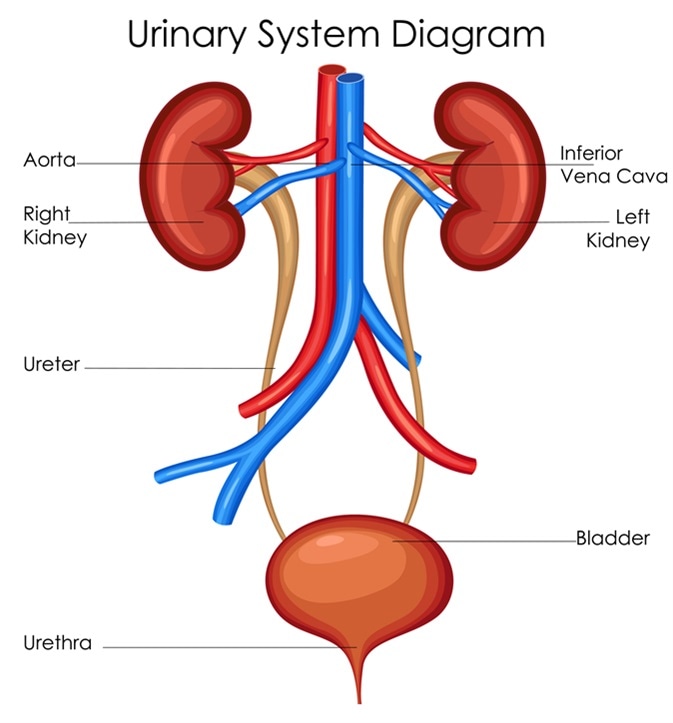
📸 Image Description: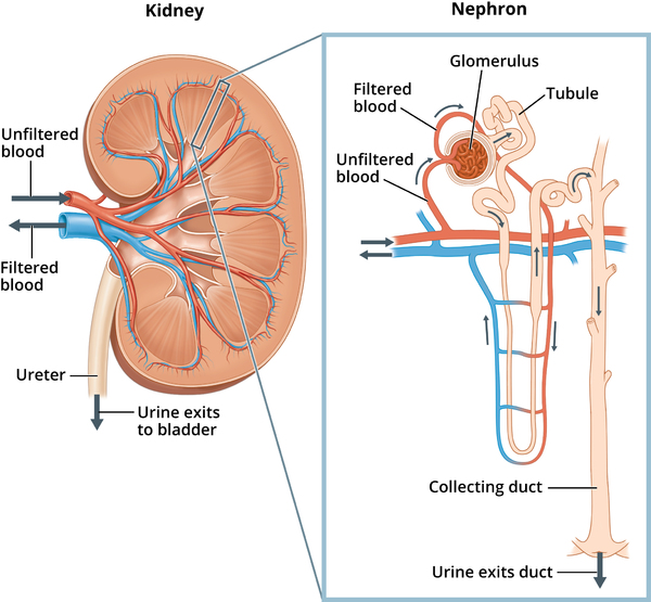
Other Urinary and Excretory Structures
- Ureters: Two narrow, muscular tubes (one from each kidney) that transport urine from the renal pelvis to the urinary bladder by peristalsis.17
- Urinary Bladder: A hollow, muscular, distensible (stretchable) organ located in the pelvic cavity that serves as a temporary storage reservoir for urine.17
- Urethra: A tube that conveys urine from the urinary bladder to the outside of the body during urination (micturition). In males, the urethra also serves as a passageway for semen. The flow is controlled by an involuntary internal sphincter and a voluntary external sphincter.17
- Other Excretory Pathways:
- Lungs: Excrete carbon dioxide and water vapor.5
- Skin: Excretes excess water, salts, and small amounts of urea in sweat.5
- Liver: Detoxifies substances and converts toxic ammonia into less toxic urea for excretion by the kidneys.5
C. Process of Urine Formation (in the Nephron)
Urine formation in the nephron involves three main processes:
- Glomerular Filtration: Occurs in the renal corpuscle. High pressure in the glomerulus forces water and small solutes from blood plasma into Bowman's capsule, forming glomerular filtrate. Large proteins and blood cells are retained in the blood.5 Approximately 180 liters of filtrate are produced per day.
- Tubular Reabsorption: As filtrate flows through the renal tubules, about 99% of its water and most useful solutes (glucose, amino acids, ions) are selectively reabsorbed back into the blood. This process is crucial to prevent the loss of vital substances. The PCT is the major site of reabsorption, while the DCT and collecting duct perform fine-tuning, regulated by hormones.5
- Tubular Secretion: The active transport of certain waste products (e.g., urea, uric acid), excess ions (H⁺, K⁺), and drugs from the blood into the filtrate within the renal tubules. This process helps eliminate wastes not filtered initially and is critical for controlling blood pH.5
The kidneys are far more than simple waste filters; they are master regulators of the body's internal environment.17 By precisely controlling the amount of water and solutes reabsorbed or secreted, they maintain the volume, osmolarity (solute concentration), and pH of the blood and other body fluids within narrow physiological limits. This homeostatic regulation is vital for the proper functioning of all body cells and systems.
🛡️ VI. The Immune System
A. Overview and Primary Functions
The immune system is a complex network of cells, tissues, and organs that work together to protect the body against pathogens—disease-causing agents such as bacteria, viruses, fungi, and parasites—as well as against abnormal body cells, such as cancer cells.20 A fundamental characteristic of the immune system is its ability to distinguish between "self" (the body's own cells and molecules) and "non-self" (foreign invaders or altered self cells).20
B. Key Cells and Organs of Immunity
Cells
White Blood Cells (Leukocytes): These are the primary effector cells of the immune system.
- Phagocytes: Cells that engulf and digest pathogens and cellular debris. Examples include Neutrophils and Macrophages.20
- Lymphocytes: Crucial for adaptive immunity.
- B-lymphocytes (B cells): Mature in bone marrow. When activated, they become plasma cells that produce antibodies (humoral immunity).20
- T-lymphocytes (T cells): Mature in the thymus. Responsible for cell-mediated immunity. Key types are Helper T cells (CD4+), which orchestrate the immune response, and Cytotoxic T cells (CD8+), which kill infected cells.20
- Natural Killer (NK) cells: A type of lymphocyte involved in innate immunity; they can kill certain tumor cells and virus-infected cells without prior sensitization.
Organs
- Primary Lymphoid Organs: Sites of lymphocyte production and maturation. Include the Bone Marrow (where all blood cells originate and B cells mature) and the Thymus (where T cells mature).20
- Secondary Lymphoid Organs and Tissues: Sites where mature lymphocytes encounter antigens. Include the Spleen, Lymph Nodes, Tonsils, and Mucosa-Associated Lymphoid Tissue (MALT).21
C. Innate (Nonspecific) Immunity vs. Adaptive (Specific) Immunity
The immune system has two major, interconnected branches:
Innate Immunity (Nonspecific Defense)
- The body's first line of defense, present from birth.20
- Provides an immediate, rapid, and nonspecific response.
- Lacks immunological memory.20
- Components include: Physical barriers (skin, mucus), chemical barriers (stomach acid, lysozyme), cellular defenses (phagocytes, NK cells), soluble factors (complement system, interferons), and inflammation.20
Adaptive Immunity (Specific Defense or Acquired Immunity)
- The body's second line of defense, develops after exposure to a specific pathogen.20
- Slower initial response, but is highly specific.
- Exhibits immunological memory, leading to a faster, stronger secondary response. This is the principle behind vaccination.20
- Mediated by lymphocytes and has two branches:
- Humoral Immunity: Involves B cells and antibodies, effective against extracellular pathogens.20
- Cell-Mediated Immunity: Involves T cells (Helper and Cytotoxic), effective against intracellular pathogens and cancer cells.20
Table 3: Comparison of Innate and Adaptive Immunity
| Feature | Innate Immunity | Adaptive Immunity |
|---|---|---|
| Specificity | Nonspecific (recognizes general pathogen features) | Highly specific (recognizes particular antigens) |
| Response Time | Rapid (minutes to hours) | Slower initial response (days); faster secondary response |
| Memory | No immunological memory | Immunological memory present |
| Key Cells | Phagocytes (macrophages, neutrophils), NK cells | Lymphocytes (B cells, T cells) |
| Key Molecules | Complement, interferons, cytokines | Antibodies, cytokines |
D. Antigens, Antibodies, and Their Interaction
Antigen (Ag): Any molecule that can be recognized by the immune system and elicit an immune response. The specific part of an antigen that is recognized is called an epitope.20
Antibody (Ab) / Immunoglobulin (Ig): Y-shaped proteins produced by plasma cells. Each antibody has at least two identical antigen-binding sites that are structurally complementary to a specific epitope, allowing for high-specificity binding.20
Functions of Antibodies:
- Neutralization: Bind to toxins or viruses, preventing them from harming cells.
- Opsonization: Coat pathogens, enhancing phagocytosis.
- Agglutination/Precipitation: Clump pathogens together for easier removal.
- Complement Activation: Triggers the complement system, leading to pathogen lysis.
📸 Image Description:
The concept of immunological memory, a hallmark of adaptive immunity, forms the scientific basis for vaccination.20 Vaccines introduce harmless forms of antigens into the body, triggering a primary adaptive immune response and generating long-lived memory B and T cells. If later exposed to the actual pathogen, these memory cells mount a swift and robust secondary immune response that typically prevents or lessens the disease.
🦋 VII. The Endocrine System
A. Overview and Primary Functions
The endocrine system is a complex network of ductless glands that produce and secrete chemical messengers called hormones directly into the bloodstream.2 These hormones travel to target cells or organs, which possess specific receptors that recognize them, initiating a response.2
The endocrine system regulates a wide array of slow, long-term processes, including growth, metabolism, mood, reproduction, and stress responses, playing a key role in homeostasis.2, 23
B. Major Endocrine Glands, Hormones, and Their Functions
📸 Image Description:
Table 4: Major Endocrine Glands and Their Hormones
| Gland | Hormone(s) Secreted | Main Function(s) |
|---|---|---|
| Hypothalamus | Releasing & Inhibiting Hormones | Controls release of hormones from anterior pituitary2 |
| Antidiuretic Hormone (ADH) (Vasopressin) | Promotes water reabsorption by kidneys24 | |
| Oxytocin | Stimulates uterine contractions and milk ejection3 | |
| ADH & Oxytocin are produced in hypothalamus, released from posterior pituitary. | ||
| Pituitary Gland | Anterior: GH, Prolactin, ACTH, TSH, FSH, LH | Regulates growth, milk production, stress response, metabolism, and reproduction by targeting other glands24 |
| Posterior: ADH, Oxytocin (storage & release) | As described above | |
| Thyroid Gland | Thyroxine (T4) & Triiodothyronine (T3); Calcitonin | Regulates metabolism (T3/T4); Lowers blood calcium (Calcitonin)2 |
| Parathyroid Glands | Parathyroid Hormone (PTH) | Increases blood calcium levels2 |
| Adrenal Glands | Cortex: Glucocorticoids (Cortisol), Mineralocorticoids (Aldosterone) | Regulates stress response, blood glucose, and inflammation (Cortisol); Regulates electrolyte/water balance (Aldosterone)2, 24 |
| Medulla: Epinephrine & Norepinephrine | Mediates "fight-or-flight" response23 | |
| Pancreas | Insulin; Glucagon | Lowers blood glucose (Insulin); Raises blood glucose (Glucagon)24 |
| Pineal Gland | Melatonin | Regulates sleep-wake cycles (circadian rhythms)2 |
| Ovaries (Females) | Estrogens; Progesterone | Regulates female reproductive cycle and secondary sexual characteristics24 |
| Testes (Males) | Testosterone | Regulates sperm production and male secondary sexual characteristics24 |
C. Hormone Action Mechanisms (Briefly)
- Steroid Hormones and Thyroid Hormones: Lipid-soluble, diffuse across the cell membrane, bind to intracellular receptors, and alter gene expression directly. Effects are slower but longer-lasting.
- Peptide/Protein Hormones and Catecholamines: Water-soluble, cannot cross the cell membrane, bind to surface receptors. This activates intracellular second messengers (like cAMP), leading to a rapid but shorter-lived cellular response.
A key feature of endocrine regulation is the hypothalamus-pituitary axis, a central control system.2 The hypothalamus controls the pituitary gland, which in turn directs other endocrine glands. Furthermore, many endocrine pathways are regulated by negative feedback loops, where the final hormone product inhibits its own production pathway. This self-regulating mechanism is essential for maintaining hormone levels within an optimal range for homeostasis.2
🧠 VIII. The Nervous System
A. Overview and Primary Functions
The nervous system is the body's primary rapid communication, control, and coordination system.3 It detects stimuli (sensory input), processes information (integration), and elicits responses (motor output).27 It governs our thoughts, emotions, memory, and control of movement.
B. The Neuron: Structure and Function
The neuron (nerve cell) is the basic unit of the nervous system, specialized for transmitting nerve impulses.27
- Cell Body (Soma): Contains the nucleus and is the metabolic center.
- Dendrites: Branched extensions that receive signals from other neurons.
- Axon: A single, long projection that transmits nerve impulses away from the cell body. It may be covered by an insulating myelin sheath (formed by Schwann cells in the PNS or oligodendrocytes in the CNS), which speeds up conduction. Gaps in the myelin are called Nodes of Ranvier.
- Axon Terminals: Branched endings of an axon that form junctions (synapses) with other cells.
📸 Image Description:
C. Nerve Impulse (Action Potential)
An action potential is a rapid, self-propagating reversal of electrical potential across the neuron's membrane. It is an "all-or-none" event triggered when a stimulus depolarizes the membrane to a threshold potential (around -55 mV).
Process:
- Depolarization: Voltage-gated Na⁺ channels open, and Na⁺ rushes into the cell, making the inside positive.
- Repolarization: Na⁺ channels inactivate, and voltage-gated K⁺ channels open. K⁺ rushes out, restoring the negative charge.
- Hyperpolarization: K⁺ channels may remain open briefly, causing an "undershoot".
- The sodium-potassium pump restores the original ion distributions.
In myelinated axons, action potentials "jump" between Nodes of Ranvier (saltatory conduction), which is much faster.
D. Synaptic Transmission
A synapse is the junction where a neuron communicates with another cell, typically using chemical messengers called neurotransmitters.27
Process at a Chemical Synapse:
- Action potential arrives at the presynaptic axon terminal.
- Voltage-gated Ca²⁺ channels open, and Ca²⁺ enters the terminal.
- Ca²⁺ influx triggers the release of neurotransmitters from synaptic vesicles into the synaptic cleft.
- Neurotransmitters bind to receptors on the postsynaptic membrane, causing a postsynaptic potential.
- Excitatory Postsynaptic Potential (EPSP) makes the neuron more likely to fire.
- Inhibitory Postsynaptic Potential (IPSP) makes the neuron less likely to fire.
- Neurotransmitters are quickly removed by enzymatic degradation, reuptake, or diffusion.
E. Divisions of the Nervous System
- Central Nervous System (CNS): Consists of the brain and spinal cord. It is the primary integration and command center.27 Major brain parts include the Cerebrum, Cerebellum, Brainstem, and Diencephalon.
- Peripheral Nervous System (PNS): Consists of all nervous tissue outside the CNS (nerves and ganglia). It connects the CNS to the body.27
- Sensory (Afferent) Division: Transmits sensory information to the CNS.
- Motor (Efferent) Division: Transmits motor commands from the CNS. It is subdivided into:
- Somatic Nervous System (SNS): Controls voluntary movements (skeletal muscles).3
- Autonomic Nervous System (ANS): Controls involuntary functions (smooth muscle, cardiac muscle, glands).3 It has two branches: the Sympathetic division ("fight-or-flight") and the Parasympathetic division ("rest-and-digest").
📸 Image Description:
A fundamental unit of nervous system response is the reflex arc, which mediates a reflex—a rapid, involuntary, and predictable response to a specific stimulus.27 Reflexes allow for extremely fast protective actions because the signal does not need to travel to the brain for conscious processing before a response is initiated.
👀 IX. Sensory Organs
A. Overview of Sensation
Sensation is the process by which sensory receptors detect stimuli and convert this energy into electrical signals (transduction). These signals are sent to the CNS for processing and perception (conscious awareness).31
B. The Eye: Structure and Function in Vision
- Sclera: The tough, white outer layer. The transparent front part is the Cornea, which is the primary light refractor.31
- Choroid: The pigmented, vascular middle layer that nourishes the retina and absorbs stray light.31
- Iris: The colored, muscular part that controls the size of the Pupil, regulating light entry.31
- Lens: A flexible structure that focuses light onto the retina. Its shape is changed by the ciliary muscle for focusing at different distances (accommodation).31
- Retina: The light-sensitive inner layer containing photoreceptors.31
- Rods: For night vision (scotopic), detect shades of gray. Highly sensitive to low light.
- Cones: For color vision (photopic) and sharp detail. Concentrated in the fovea, the area of highest visual acuity.33
- Optic Nerve: Transmits visual information from the retina to the brain.31 The point where it leaves the eye is the "blind spot".
📸 Image Description:
C. The Ear: Structure and Function in Hearing and Balance
The ear has two functions: hearing and balance (equilibrium).31
- Outer Ear: Collects sound waves. Consists of the Pinna (auricle), External Auditory Canal, and Tympanic Membrane (eardrum), which vibrates in response to sound.35
- Middle Ear: An air-filled cavity that transmits and amplifies vibrations. Contains three tiny bones (ossicles): the Malleus (hammer), Incus (anvil), and Stapes (stirrup). The Eustachian tube equalizes pressure.35
- Inner Ear: A fluid-filled labyrinth containing sensory receptors.
- Cochlea (for Hearing): A spiral-shaped chamber containing the Organ of Corti, which has hair cells (mechanoreceptors). Vibrations cause these hair cells to bend, generating nerve impulses for hearing.31
- Vestibular System (for Balance): Consists of three semicircular canals (detecting rotational movement)32 and two otolith organs (the utricle and saccule, detecting linear acceleration and gravity). Both use hair cells to detect fluid or crystal movement.
📸 Image Description:
D. Briefly: Taste (Gustation), Smell (Olfaction), and Touch (Somatosensation)
- Taste (Gustation): Detected by chemoreceptors in taste buds on the tongue, which recognize sweet, sour, salty, bitter, and umami qualities.31
- Smell (Olfaction): Detected by chemoreceptors in the olfactory epithelium in the nasal cavity. Smell is closely linked to taste.31
- Touch (Somatosensation): Includes sensations from the skin detected by various receptors: Mechanoreceptors (pressure, touch), Thermoreceptors (temperature), and Nociceptors (pain).32
A unifying principle across all sensory systems is transduction: the conversion of a specific type of stimulus energy (light, sound, chemical, pressure) into an electrical signal that the nervous system can process.31 The ear is an excellent example of a dual-role organ, housing structures for both hearing (cochlea) and balance (vestibular system), both of which rely on mechanoreceptor hair cells.31
🦴 X. The Musculoskeletal System
A. Overview and Primary Functions
The musculoskeletal system is an integrated system of bones, muscles, cartilage, tendons, and ligaments. Its functions include support, protection, movement, mineral storage (calcium), and blood cell production (hematopoiesis).37
B. The Skeletal System
Bone Structure
- Types of Bone Tissue:
- Compact Bone: The dense, hard outer layer organized into osteons. Provides strength and rigidity.37
- Spongy Bone: A porous inner layer with a honeycomb-like structure of trabeculae. The spaces are filled with red bone marrow for hematopoiesis.37
- Anatomy of a Long Bone: Consists of a shaft (diaphysis) and ends (epiphyses). Covered by a membrane called the periosteum. The central medullary cavity contains yellow bone marrow (fat storage) in adults.38
- Bone Cells:
- Osteoblasts: Bone-forming cells ("Build").37
- Osteoclasts: Bone-resorbing cells ("Crush").37
- Osteocytes: Mature bone cells that maintain the bone matrix.
Divisions of the Skeleton
- Axial Skeleton: The central axis, including the skull, vertebral column, and rib cage.37
- Appendicular Skeleton: The limbs and the pectoral (shoulder) and pelvic girdles that attach them to the axial skeleton.37
📸 Image Description:
C. The Muscular System
Types of Muscle Tissue
- Skeletal Muscle: Attached to bones, responsible for voluntary movement. It is striated.37
- Smooth Muscle: Found in walls of hollow organs, responsible for involuntary movements. It is non-striated.
- Cardiac Muscle: Found only in the heart, responsible for involuntary pumping. It is striated and has intercalated discs.
D. Mechanism of Muscle Contraction (Sliding Filament Theory)
Muscle contraction occurs when the basic contractile units, called sarcomeres, shorten.
Sarcomere Structure
The sarcomere is the region between two Z-lines.
- Thin Filaments: Made of actin and regulatory proteins (tropomyosin and troponin).
- Thick Filaments: Made of myosin, with heads that can bind to actin.
📸 Image Description: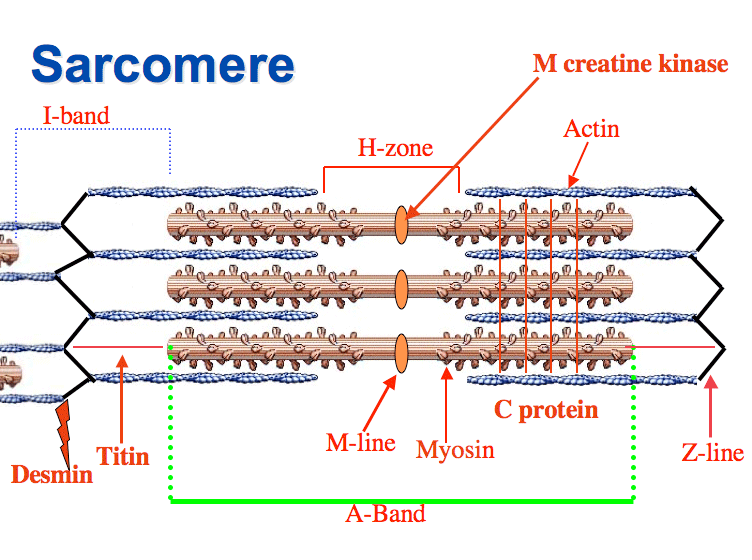
Sliding Filament Process
- Excitation: A motor neuron releases acetylcholine (ACh) at the neuromuscular junction, generating a muscle action potential.37
- Calcium Release: The action potential travels down T-tubules, causing the sarcoplasmic reticulum to release Ca²⁺ ions.
- Cross-Bridge Formation: Ca²⁺ binds to troponin, causing tropomyosin to move and expose the myosin-binding sites on actin. Myosin heads then bind to actin, forming cross-bridges.
- Power Stroke: The myosin heads pivot, pulling the thin filaments toward the center of the sarcomere. ATP is required to detach the myosin heads and "recock" them for the next cycle.
- Relaxation: When nerve stimulation ends, Ca²⁺ is pumped back into the sarcoplasmic reticulum, tropomyosin covers the actin binding sites, and the muscle fiber relaxes.